
🇺🇸🇬🇧 Menu of this website's pages IN ENGLISH :


Thank you for SHARING this webpage via:
Teratoma of Mediastinum
PreOp investigations revealed a marked elevation of the Ca 19.9* cancer antigen ( patient's serum level of Ca 19.9 was 101.1 U/mL, instead of < 37.0 )
VIDEO :
Photographs :
PreOp CXRs. There was a slight elevation of the right hemidiaphragm.
Figures 3–6:
PreOp CTs
Figures 7–12:
IntraOp findings, as via a right Limited thoracotomy through the bed of the 5th rib (i.e. the 4th interspace). The teratoma was abutting the right phrenic nerve along its course on the pericardium; the nerve was dissected free from the tumour and spared.
PostOp:
Histopathology findings: a benign, mature cystic teratoma, measuring 5.2 cm (maximal dimension) and arising from thymus. There were elements of skin and adipose tissue as well as sebaceous glands, columnar ciliated epithelium as well as islets of pancreas* tissue and keratin inside the tumour. There were focal degenerative changes. There were no abnormalities within the thymus specimen, measuring 11 cm.
* The presence of islets of pancreas tissue within the resected tumour interprets the preoperatively elevated Ca 19.9 levels on this case (Ca 19.9 was postoperatively measured within the normal range)
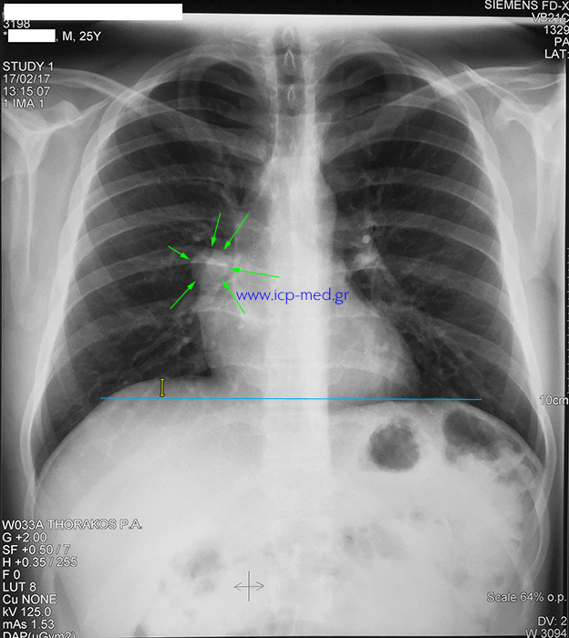
1. Preoperative CXR (posteroanterior) of the incidental finding. The teratoma is shown by GREEN arrows. There is minimal elevation of the right hemidiaphragm.
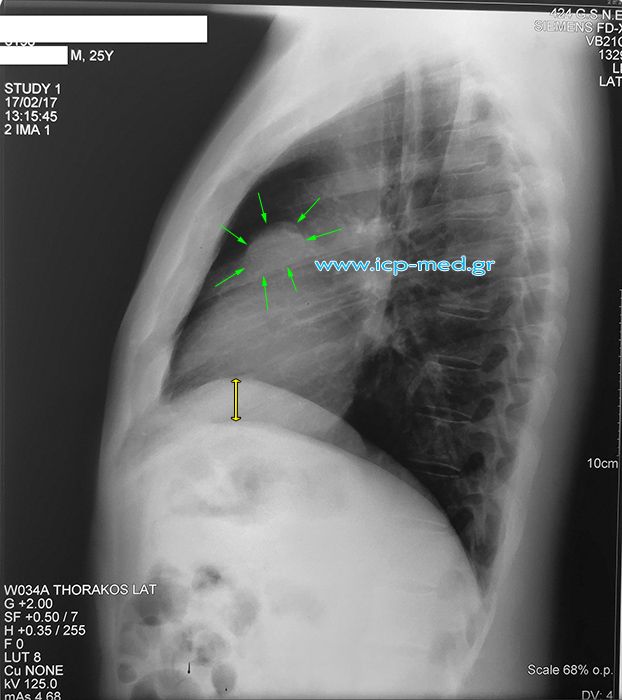
2. Preoperative CXR (lateral) of the incidental finding. The teratoma is shown by GREEN arrows.
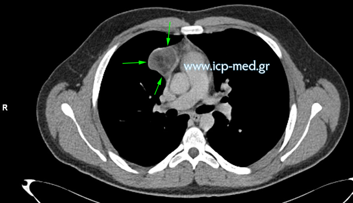
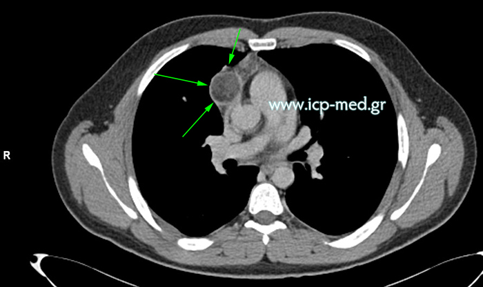
3. Preoperative CT (mediastinal window). GREEN arrows show the teratoma
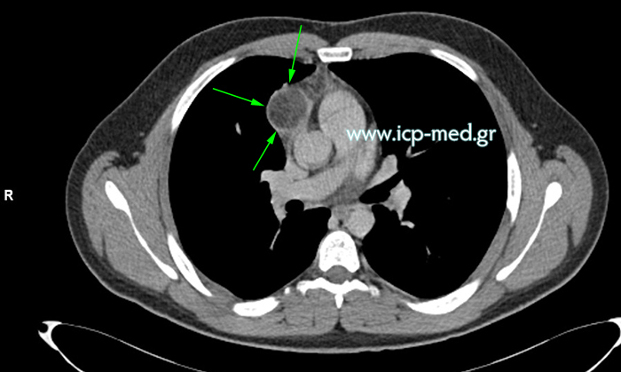
4. Preoperative CT (mediastinal window). GREEN arrows show the teratoma
5. Preoperative CT (mediastinal window). GREEN arrows show the teratoma
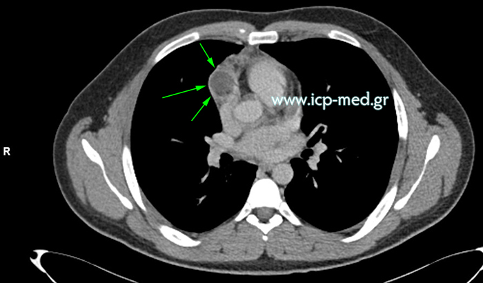
6. Preoperative CT (mediastinal window). GREEN arrows show the teratoma
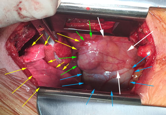
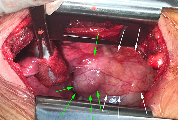
7. Intraoperative findings
via a right limited thoracotomy through the bed of the 5th rib (i.e. the 4th interspace) :- GREEN arrows show the tumour (the teratoma)
- WHITE arrows show remnants of Thymus
- YELLOW arrows show the right lung, partly deflated and retracted towards the right side
- BLUE arrows show the pericardium
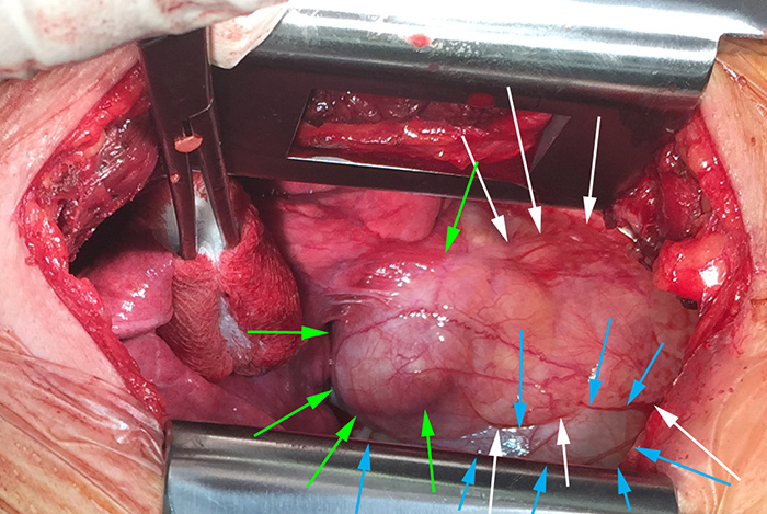
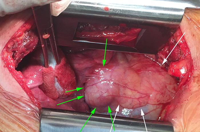
8. Intraoperative findings
via a right limited thoracotomy.
Same colours as used on photo № 7 :- GREEN arrows show the tumour (the teratoma)
- WHITE arrows show remnants of Thymus
- BLUE arrows show the pericardium
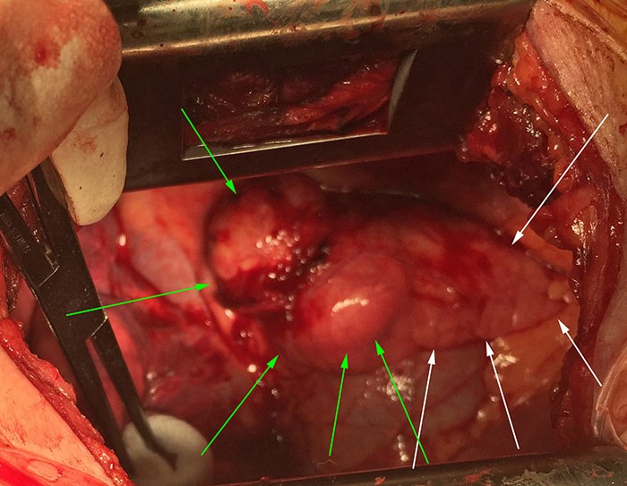
9. Intraoperative findings
via a right limited thoracotomy.
Same colours as used on photo № 7 :- GREEN arrows show the tumour (the teratoma)
- WHITE arrows show remnants of Thymus
10. Intraoperative findings
via a right limited thoracotomy.
Same colours as used on photo № 7 :- GREEN arrows show the tumour (the teratoma)
- WHITE arrows show remnants of Thymus
11. Intraoperative findings
via a right limited thoracotomy.
Same colours as used on photo № 7 :- GREEN arrows show the tumour (the teratoma)
- WHITE arrows show remnants of Thymus
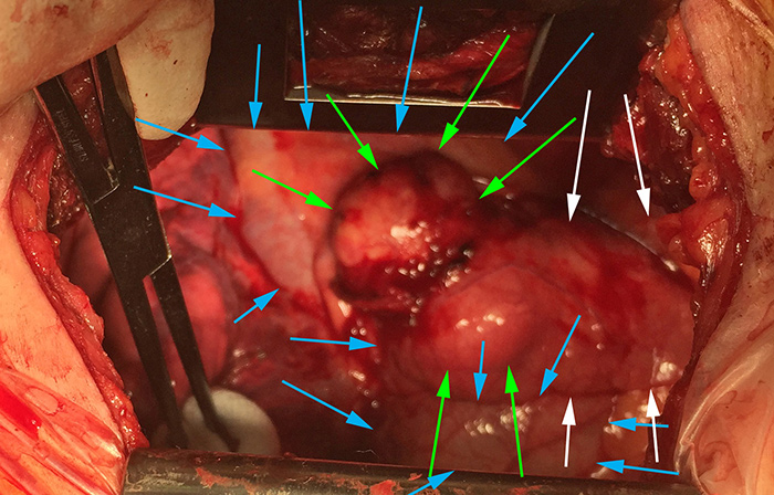
12. Intraoperative findings
via a right limited thoracotomy.
Same colours as used on photo № 7 :- GREEN arrows show the tumour (the teratoma)
- WHITE arrows show remnants of Thymus
- BLUE arrows show the pericardium
Thank you for SHARING this webpage via:

An additional MENU can be revealed by clicking on the PLUS symbol ( top – right ) ; it leads to the entirety of all English pages of this site to support the navigation.
On the contrary, the “Main” horizontal menu ( top – centre ) only links to sub–pages, but not to their children.

You might also want to browse the Writer's European site of mine.