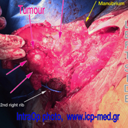
The tumour was extending along the 2nd costo–sternal joint (where a secondary mass measured 2.9 × 2.0 cm). Bone scintigraphy had preop detected increased uptake at both clavicles (their sternal ends), the manubrium and the upper part of corpus sterni, the 3rd right costal cartilage and the left 2 superior ones. The tumour abutted the ascending aorta, pericardium and SVC; it was resected along with all overlying musculocutaneous structures.
Chest wall reconstruction was carried out by using 3 tubes (filled in by methyl-methacrylate and reinforced by steel wires) and by implanting a mesh. The complex prosthesis was covered by colleagues plastic surgeons, who used a right latissimus dorsi musculocutaneous flap.
The patient was discharged home on 14th post day; he was referred for post op radiotherapy, as indicated in cases of recurrence.
Fourteen yrs before, the patient had elsewhere undergone resection of his 1st right costo-sternal joint by another colleague, who had disarticulated the sterno-clavicular and then sewn it with a steel wire (green arrows in preop images). Histopathology report had failed to identify a chondrosarcoma diagnosis at that old time!
IMAGES 2–6: Preop CXR & CTs,
IMAGES 7–11: Intraop photographs,
IMAGES 12–14: Postop result.
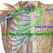
1. Line of dividing the bony structures of anterior chest wall
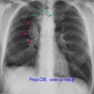
2. PreOp CXR. Magenta arrows: the tumour. Green arrows: steel wire elsewhere 14 yrs before
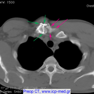
3. PreOp CT. Magenta arrows: the tumour. Green: steel wire elsewhere inserted 14 yrs before
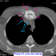
4. PreOp CT. The tumour (magenta arrows) abuts the Superior Vena Cava (cyan arrows)
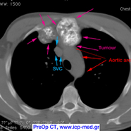
5. PreOp CT. The tumour (magenta arrows) abuts the aortic arch / ascending aorta (red arrows)
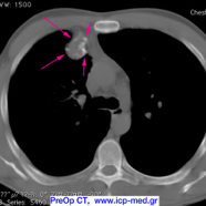
6. PreOp CT. The timour (magenta arrows), extending along the 2nd right rib
7. IntraOp photo: The left superior ribs were first divided, then the sternum was divided caudal to the right 4th interspace & the left 3rd.
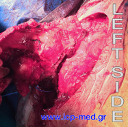
8. IntraOp photo. Another view of the internal aspect of the specimen (being further mobilised), from the left side
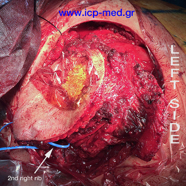
9. IntraOp photo. External view of the specimen, almost completely mobilised along with the overlying musculocutaneous structures
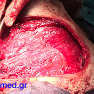
10. IntraOp photo. A right latissimus dorsi flap is sewn, to cover the underlying complex reconstruction of the chest wall.
11. IntraOp photo. View of the chest wall after the resectional procedure & the reconstruction
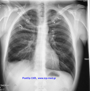
12. PostOp CXR (posteroanterior). Apparent methyl-methacrylate tubes, reinforced by steel wires at the anterior chest wall
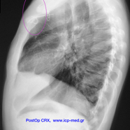
13. PostOp CXR (lateral). One can see the area of resection highlighted
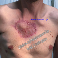
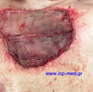
14. Appearance of the patient's anterior chest wall, fully healed.