
Mediastinal Seminoma, primary
Seminoma, primarily located in the Mediastinum (i.e. Not a metastasis from testicular tumour) was an incidental radiological finding in a 31-yo male non-smoker's case during check-up.
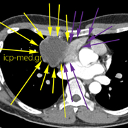
The malignant, infiltrative tumour (YELLOW arrows) was Large (measuring 8.9 cm in maximal dimension) and its resection through median sternotomy was Difficult, because it was Densely ADHERED to the following major anatomical structures (e.g. cavities of the heart etc.):
Superior Vena Cava (BLUE arrows),
Ascending Aorta (RED arrows),
Right Pulmonary Artery (GREEN arrows)
Right Atrium (cavity of the heart, dark VIOLET arrows)
Left Ventricle (cavity of the heart, MAGENTA or pink arrows)
Right Pulmonary Vein (BROWN arrows)
IMAGE 1: PreOp Chest X-Ray
IMAGES 2-6: PreOp chest CT scans
IMAGES 7-8: Specimen during and after its resection
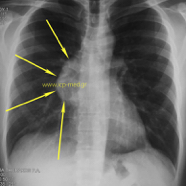
1.Preop CXR: the Seminoma appears as Confluent to the heart silhouette (right side)
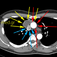
2.CT: the Seminoma abuts the SVC & the Ascending Aorta
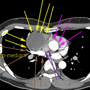
4.CT: Seminoma abuts Cavities of the heart (Right Atrium & Left Ventricle) & a right Pulmonary Vein
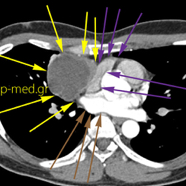
5.CT: Seminoma abuts the Rigt Atrium (cavity of heart) & a Pulmonary Vein
6.CT: Seminoma abuts teh Right Atrium (cavity of the heart)
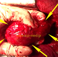
7.Specimen still in situ, moments prior to its resection
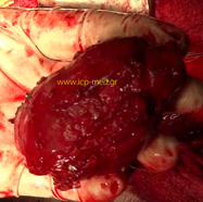
8.Specimen of Seminoma resected (size comparison against operator's fingers)