
Chest Wall Myofibroblastic Sarcoma - VATS
A Chest Wall malignant myofibroblastic Sarcoma, rapidly progressing, was presented as an incidental radiologic finding in a completely Asymptomatic 21-yo male smoker (a fit athlete)
IMAGES 1-2: Pre-op CXRs of the asymptomatic patient upon his admission.
IMAGE 3: No abnormality had been detected on another CXR, taken 14 months prior to admission
IMAGES 4-5: Pre-op CT (Computed axial Tomography) scans
IMAGES 6-9: Pre-Op MRI (Magnetic Resonance Imaging) scans
IMAGES 10-12: Intra-operative photographs of the intrathoracic tumour (VATS), then during the radical resectional procedure (Chest Wall Resection) and, finally, of the specimen resected.
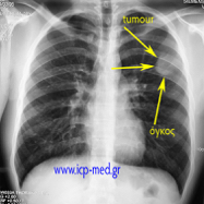
1. Preop CXR (posteroanterior) upon admission
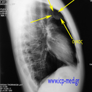
2. Preop CXR (lateral) upon admission
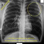
3. No abnormality detected on CXR 14 months prior to admission
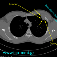
4. Preop CT scan (cyan arrows: the contact between the tumour & the 3rd rib)
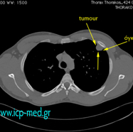
5. Preop CT scan
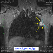
6. Preop MRI scan (coronal view; cyan arrows: the contact between the tumour & the 3rd rib)
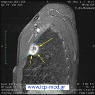
7. Preop MRI scan (sagittal view; cyan arrows: the contact between the tumour & the 3rd rib)
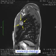
8. Preop MRI scan (sagittal view)
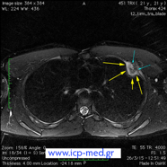
9. Preop MRI scan (axial view; cyan arrows: the contact between the tumour & the 3rd rib)
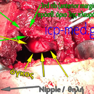
11. Intraop photo of the redical resectional procedure (Chest Wall Resection)
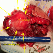
12. The finally resected specimen of the tumour along with its overlying muscular & skin tissues involved.