
LAM: Lymphangioleiomyomatosis (Lymphangiomyomatosis)
IMAGES 1-3: Multiple, thin-walled bullae (Yellow arrows) bilaterally. Also small-sized pneumothorax, left-sided (Blue arrows) & surgical emphysema in a 25-yo female non-smoker's case who had undergone lung biopsy & pleurodesis in 2008. Then, she started being administered a Sirolimus or Rapamycin treatment: more than 7 years later, she has remained Alive and with her Respiratory Function Tests IMPROVED. There has been No recurrence of pneumothorax.
IMAGES 4-11: Numerous Large bullae (Yellow arrows) in a 55-yo female post-menopausal & non-smoker's case (history of previous hysterectomy because of leiomyomas, farmer). She underwent lung biopsy on the left side.
Histopathology diagnosis of LAM was achieved in both cases and that was confirmed by HMB45 immunohistochemistry.
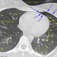
1. CT: thin-walled bullae, pneumothorax (left-sided) & surg. emphysema in a 25-yo ♀
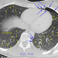
2. CT: thin-walled bullae, pneumothorax (left-sided) & surg. emphysema in a 25-yo ♀
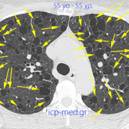
5. CT: numerous Large bullae in a 55-yo post-menopausal♀
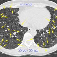
6. CT: numerous Large bullae in a 55-yo post-menopausal♀
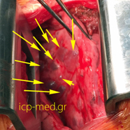
8. IntraOp photogr. of multiple bullae (Yellow arr.) in a 55-yo ♀
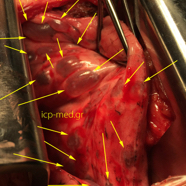
9. IntraOp photogr. of multiple bullae (Yellow arr.) in a 55-yo ♀
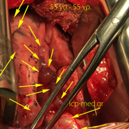
10. IntraOp photogr. of multiple bullae (Yellow arr.) in a 55-yo ♀
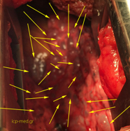
11. IntraOp photogr. of multiple bullae (Yellow arr.) in a 55-yo ♀