
Lung Foreign bodies (pulmonary & endobronchial)
FIGURES 1-2:
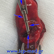
Lead shot or pellet in a 22-yo male smoker’s case:
BLUE arrows: Intrapulmonary airgun shot, made of toxic metal (Pb-Lead), removed. The long-term presence of the lead shot inside the lung (over 2 yrs) had chemically destroyed the neighbouring lung tisuue: A small-sized piece of lung had to be wedge-resected: Specimen measuring 1.5 cm (YELLOW arrows), from the “apical lower” segment of the LLL.
The scar of the pellet’s entrance would is marked by a BLUE circle.
FIGURES 3-8:
A dentist’s metallic instrument was aspirated in a 31-yo male non-smoker’s case. Endobronchial location of the foreign body inside a sub-segmental bronchus of the posterior basal segment of the LLL. Removal of the foreign body was repeatedly (5 times), yet unseccesfully, attempted by bronchoscopy; then, bronchoscopic attempts had to be abandoned for progressing expectoration of blood-stained sputum commenced.
FIG. 3-4:
Preoperative Chest X–rays (posteroanterior and left lateral)
FIG. 5-8:
Wedge resection specimen. The cylindrical–shaped foreign body is retrieved by in vitro transecting the specimen
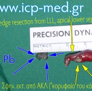
1. Lead shot or pellet

2. Scar of the entrance wound

3. Preop CXR (posteroanterior)
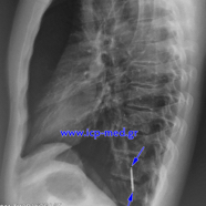
4. Preop CXR (left lateral)
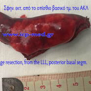
5. Specimen of Wedge Resection from the LLL
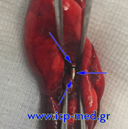
6. In vitro transection of the specimen: foreign body apparent inside
7. Retrieval of the foreign body
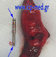
8. Pulmonary specimen and the removed foreign body