
Macleod syndrome (or Swyer–James)
Idiopathic hyperlucent lung syndrome, that had elsewhere been misdiagnosed as ‘pneumothorax, left–sided.’
IMAGE 1: A 23–yo non–smoker had a chest x–ray for ‘Occupational Health Dept.’ reasons (upon expiration of employment contract).
IMAGE 2: The subject had been absolutely Asymptomatic until the CXR was taken; he underwent, however, a chest drain insertion ‘Urgently’ elsewhere (the Non–apical drain was inserted at a small, rural,remote hospital and this insertion possibly caused penumothorax, after all).
The patient was afterwards transferred to a thoracic surgical unit because no pulmonary re–expansion had been achieved.
IMAGES 3-4: Macleod's syndrome was finally diagnosed there (with stenosis of the left pulmonary artery) along with co–existence of a pneumothorax (possibly an iatrogenic one).
IMAGE 5: New, apical chest drain led to full pulmonary re–expansion.
IMAGE 6: ‘Under–penetrating,’ expirational CXR upon discharge home
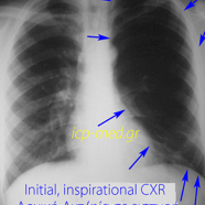
1. Initial CXR of Asymptomatic male subject with hyperlucent lung, left-sided
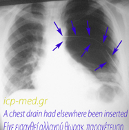
2. The non–apical chest drain, elsewhere inserted: post–procedural persistence of hyperlucency
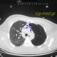
3. Chest CT: vascular desertion on the left side (left pulmonary artery stenosis) & Pneumothorax
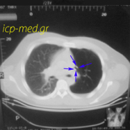
3. Chest CT: vascular desertion on the left side (left pulmonary artery stenosis) & Pneumothorax
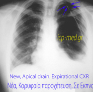
5. The new, apical chest drain inserted for treatment of the pneumothorax ‘finally caused’
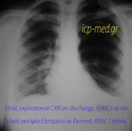
6. Final, ‘under–penetrating,’ expirational CXR upon discharge home