
IMAGES 1-6: Solid mass (max. dim. 1.7 cm, SUVmax: 12) of the right lung (RLL) in a 50-yo male smoker’s case (50 pack-yrs).
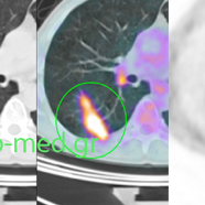
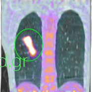
IMAGES 7-9: Solid mass (max. dim. 1.1 cm, SUVmax:7.6) of the left lung (LUL) in a 72-yo male smoker’s case (40 pack-yrs).
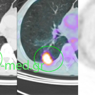
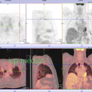
1. Preop PET/CT (Positron Emission Scanning combined with CT) in a 50-yo male smoker: solid mass of the right lung
2. Preop PET/CT (axial view) of a solid mass in the pulmonary RLL (SUVmax 12 & max. dim. 1.7 cm)
3. Preop PET/CT (coronal view) of a solid mass in the RLL, intensely hypermetabolic (SUVmax 12)
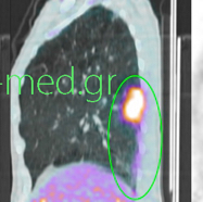
4. Preop PET/CT (sagittal veiw) of a lesion of the RLL, very suspicious for malignancy (SUVmax 12)

5.Specimen (measuring 7.5 x 5.5 x 4.8 cm) of wedge resection of the lesion, directly compared against a standard 5 ml syringe
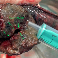
6. Specimen transected: cavity shown in its interior (pulmonary tuverculosis mimicking tumour)
7. Preop PET/CT (Positron Emission Tomography associated with CT) in a 72-yo male smoker: solid mass of the left lung
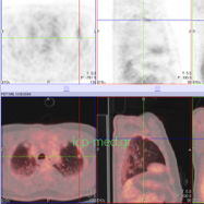
8. Preop PET/CT of the hyermetabolic lesion (measuring 1.1. cm) of the pulmonary LUL. Postop diagnosis: TBC
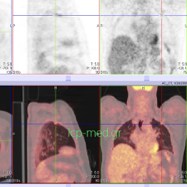
9. Preop PET/CT of the LUL lesion (measuring 1.1 cm), that was very suspicious for (mimicking) malignancy; final postop diagnosis:TBC