Chest Wall Myofibroblastic Sarcoma - VATS
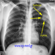
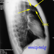
A Chest Wall malignant myofibroblastic Sarcoma, rapidly progressing, was presented as an incidental radiologic finding in a completely Asymptomatic 21-yo male smoker (a fit athlete)
IMAGES 1-2: Pre-op CXRs of the asymptomatic patient upon his admission.
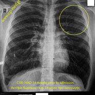
IMAGE 3: No abnormality had been detected on another CXR, taken 14 months prior to admission
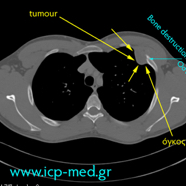
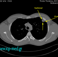
IMAGES 4-5: Pre-op CT (Computed axial Tomography) scans
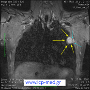
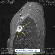
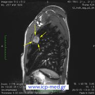
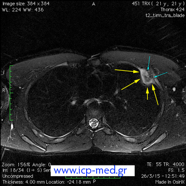
IMAGES 6-9: Pre-Op MRI (Magnetic Resonance Imaging) scans
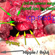
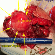
IMAGES 10-12: Intra-operative photographs of the intrathoracic tumour (VATS), then during the radical resectional procedure (Chest Wall Resection) and, finally, of the specimen resected.

