
IMAGES 7, 10: Large Hydatid Cyst of the right lung (RLL) in a 19-yo male smoker's case (2 pack-yrs): Intraoperative photographs (Prior to & Post Removal of the Cyst)
images 8-9: Sizeable Hydatid Cyst of the left lung (LLL) in a 19-yo male smoker's case (6 pack-yrs): Intraoperative photographs (Prior to & Post Removal of the Cyst)
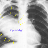
1. Preop CXR (anteroposterior): Gigantic Hydatid Cyst (18 x 12 cm, YELLOW arrows) of the right lung
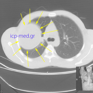
3. Preop CT: Gigantic Hydatid Cyst (18 x 12 cm, YELLOW arrows) of the right lung, abutting the hilum vessels
4. Preop CT: Gigantic Hydatid Cyst (18 x 12 cm, YELLOW arrows) of the right lung.
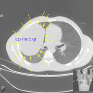
5. Preop CT: Gigantic Hydatid Cyst (18 x 12 cm, YELLOW arrows) of the right lung.
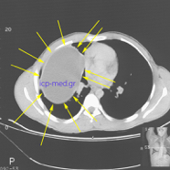
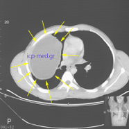
6. Preop CT: Gigantic Hydatid Cyst (18 x 12 cm, YELLOW arrows) of the right lung, abutting the hilum vessels & the heart
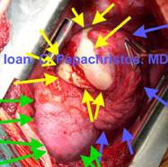
7. Intraop photograph: Sizeable Hydatid Cyst (max.dim. 6 cm, YELLOW) of the pulm. RLLL (BLUE). GREEN: right hemidiaphragm
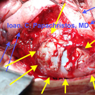
8. Intraop photograph: Large Hydatid Cyst (6.9 x 5.4 cm, YELLOW arrows) of the pulm. LLL (BLUE arrows)
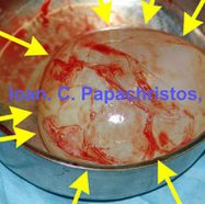
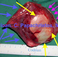
9. Large hydatid Cyst (6.9 x 5.4 cm) of the LLL, entirely enucleated (specimen)
10. Sizeable Hydatid Cyst (m.d. 6 cm, YELLOW arrows) of the RLL, wedgely resected. GREEN: wedge resection margins (specimen)