Lung Foreign bodies (pulmonary & endobronchial)
FIGURES 1-2:
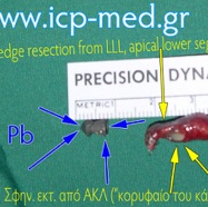
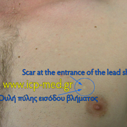
Lead shot or pellet in a 22-yo male smoker’s case:
BLUE arrows: Intrapulmonary airgun shot, made of toxic metal (Pb-Lead), removed. The long-term presence of the lead shot inside the lung (over 2 yrs) had chemically destroyed the neighbouring lung tisuue: A small-sized piece of lung had to be wedge-resected: Specimen measuring 1.5 cm (YELLOW arrows), from the “apical lower” segment of the LLL.
The scar of the pellet’s entrance would is marked by a BLUE circle.
FIGURES 3-8:
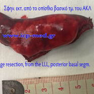
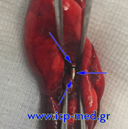
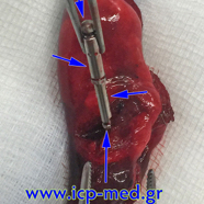
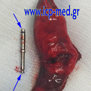
A dentist’s metallic instrument was aspirated in a 31-yo male non-smoker’s case. Endobronchial location of the foreign body inside a sub-segmental bronchus of the posterior basal segment of the LLL. Removal of the foreign body was repeatedly (5 times), yet unseccesfully, attempted by bronchoscopy; then, bronchoscopic attempts had to be abandoned for progressing expectoration of blood-stained sputum commenced.
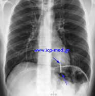
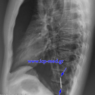
FIG. 3-4:
Preoperative Chest X–rays (posteroanterior and left lateral)
FIG. 5-8:
Wedge resection specimen. The cylindrical–shaped foreign body is retrieved by in vitro transecting the specimen
