Anterior Chest Wall Chondrosarcoma
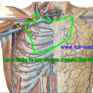
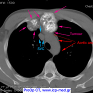
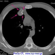
A 38-yo male, non-smoker, had a recurrent malignant chondrosarcoma (measuring 6.6 × 5.4 × 2.9 cm) located at the superior part of his sternum.
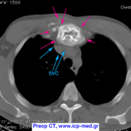
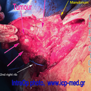
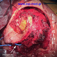
The tumour was extending along the 2nd costo–sternal joint (where a secondary mass measured 2.9 × 2.0 cm). Bone scintigraphy had preop detected increased uptake at both clavicles (their sternal ends), the manubrium and the upper part of corpus sterni, the 3rd right costal cartilage and the left 2 superior ones. The tumour abutted the ascending aorta, pericardium and SVC; it was resected along with all overlying musculocutaneous structures.
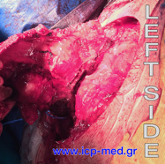
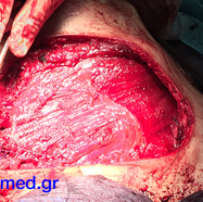
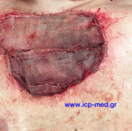
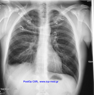
Chest wall reconstruction was carried out by using 3 tubes (filled in by methyl-methacrylate and reinforced by steel wires) and by implanting a mesh. The complex prosthesis was covered by colleagues plastic surgeons, who used a right latissimus dorsi musculocutaneous flap.
The patient was discharged home on 14th post day; he was referred for post op radiotherapy, as indicated in cases of recurrence.
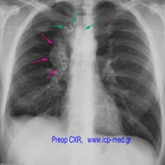
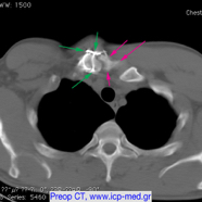
Fourteen yrs before, the patient had elsewhere undergone resection of his 1st right costo-sternal joint by another colleague, who had disarticulated the sterno-clavicular and then sewn it with a steel wire (green arrows in preop images). Histopathology report had failed to identify a chondrosarcoma diagnosis at that old time!
IMAGES 2–6: Preop CXR & CTs,
IMAGES 7–11: Intraop photographs,
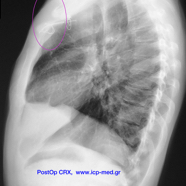
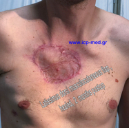
IMAGES 12–14: Postop result.
